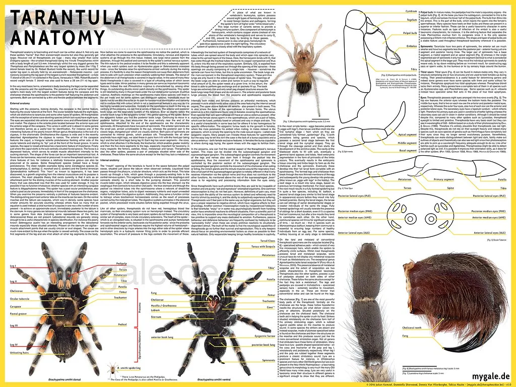
The Tarantula’s Anatomy An Overview
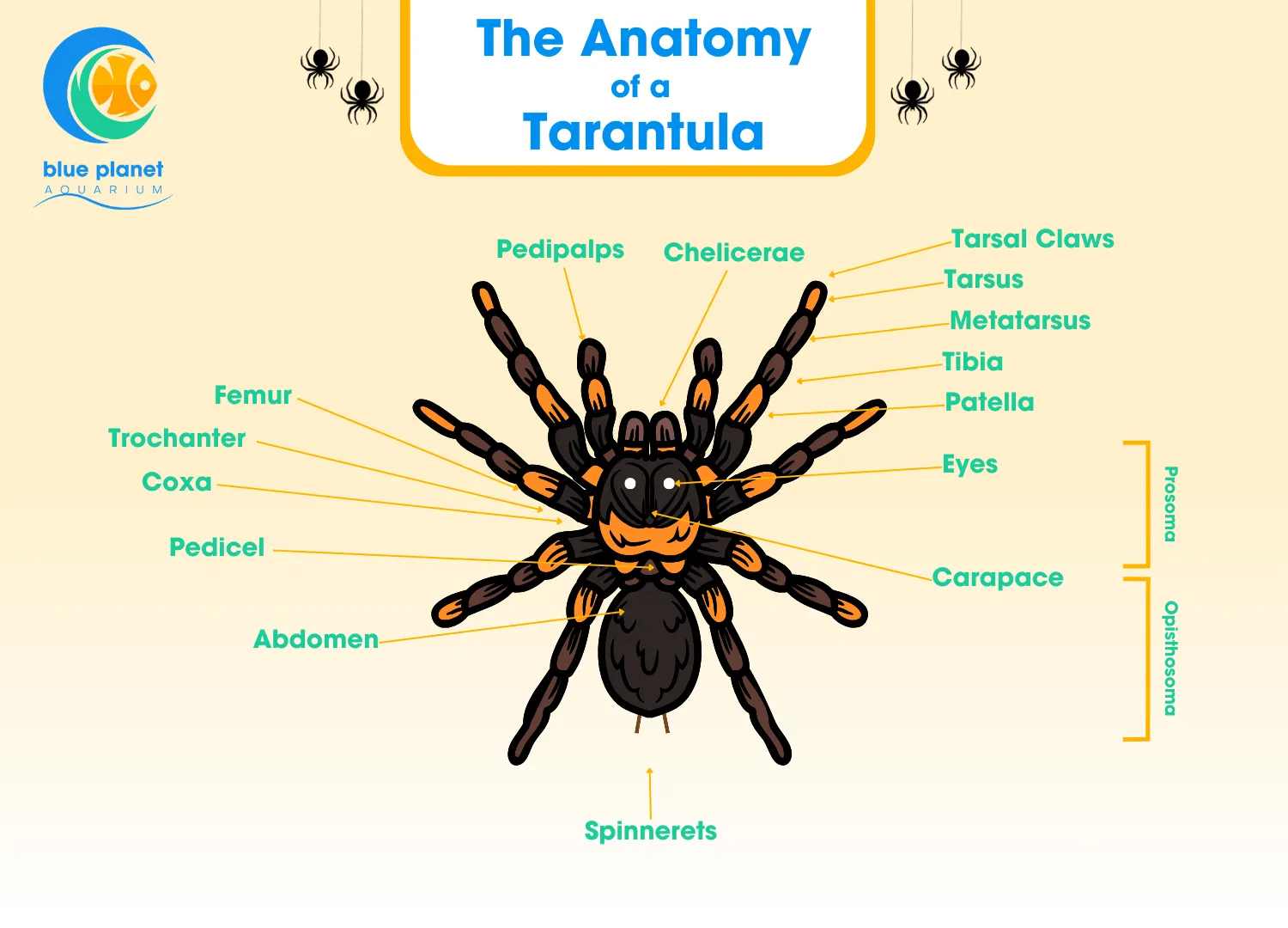
Tarantulas, the impressive arachnids captivating enthusiasts worldwide, boast a complex internal architecture that allows them to thrive in diverse environments. Their survival and behavior depend on an intricate network of specialized organs. Understanding these organs is crucial to appreciating the spider’s capacity to hunt, breathe, and perceive the world around it. From the outer exoskeleton to the inner workings of their nervous system, every part plays a critical role. This detailed overview of tarantula anatomy will help you understand how each organ contributes to its survival.
Exoskeleton and Its Significance
The exoskeleton, a tough, protective outer layer composed primarily of chitin, is a defining characteristic of all arthropods, including tarantulas. This rigid shell serves as both armor against predators and a framework for muscle attachment. The exoskeleton prevents water loss, crucial for survival in arid environments, and offers structural support. However, because it is inflexible, the exoskeleton dictates a unique life cycle stage known as molting. The exoskeleton of a tarantula is not just a simple shell; it’s a complex structure that protects and supports the spider throughout its life, allowing it to survive in various environments. The rigid structure also protects from physical harm, making tarantulas quite resilient.
Molting Process The Shedding of Old
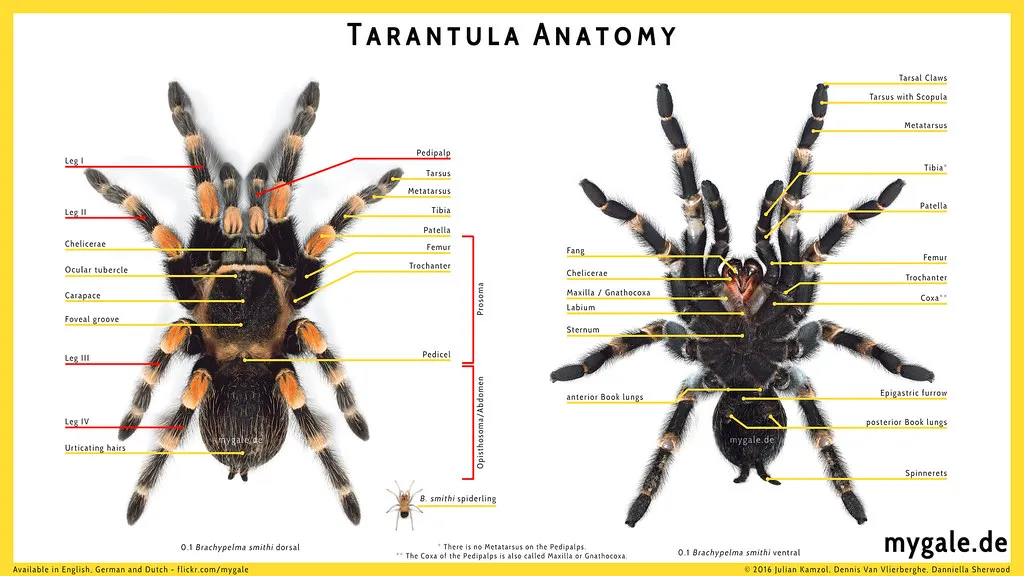
Molting is a vital process where tarantulas shed their old exoskeleton to grow and regenerate damaged limbs. This process is driven by hormones, with the spider creating a new, soft exoskeleton beneath the old one. The tarantula then absorbs water and expands, cracking open the old exoskeleton. The spider carefully pulls itself out, leaving behind its previous shell. During molting, the tarantula is incredibly vulnerable, as the new exoskeleton takes time to harden. Once molted, the spider will appear brighter in color and significantly larger. Molting is also a critical opportunity for tarantulas to regrow lost limbs. The molting process ensures continued growth and the ability to repair damage.
Respiratory System How Tarantulas Breathe
Tarantulas utilize two primary respiratory organs: book lungs and tracheae. Book lungs, unique to arachnids, are located on the underside of the abdomen and resemble the pages of a book. These structures facilitate gas exchange, with blood flowing through thin lamellae (plates) exposed to the air. Tracheae are a series of fine tubes that carry air directly to the tissues. This dual system allows for efficient oxygen uptake and carbon dioxide removal, supporting the tarantula’s active lifestyle. These organs work in concert to supply the tarantula with the oxygen it needs to function properly.
Book Lungs The Primary Breathing Organs
Book lungs are one of the primary respiratory organs in tarantulas, essential for gas exchange. Located ventrally in the abdomen, they consist of numerous thin, leaf-like lamellae that increase the surface area for oxygen absorption. The lamellae are filled with hemolymph, which allows for the diffusion of gases. Air enters through small openings called spiracles, and oxygen is absorbed into the hemolymph. Book lungs are highly efficient and allow tarantulas to extract oxygen from the air, which is crucial for their survival and activity levels. The intricate design of book lungs ensures efficient oxygen uptake, supporting the tarantula’s metabolic needs.
Tracheae and Their Role
Tracheae are another essential component of the tarantula’s respiratory system, providing an alternative pathway for oxygen delivery. These are a network of branching tubes that extend throughout the body, directly delivering oxygen to the tissues. This system supplements the book lungs, ensuring sufficient oxygen supply, especially during periods of high activity. Tracheae are particularly important in smaller tarantula species and play a crucial role in the distribution of oxygen throughout the body. This system allows for effective gas exchange and supports the tarantula’s overall metabolic rate. Tracheae’s direct delivery of oxygen ensures efficient respiration, vital for tarantula function.
Circulatory System Understanding the Tarantula’s Blood Flow
Tarantulas have an open circulatory system, where blood (hemolymph) is not always contained within vessels. Instead, hemolymph bathes the tissues, providing nutrients and removing waste. A central heart pumps hemolymph throughout the body, circulating it through the hemocoel (body cavity). This system efficiently delivers essential substances to the tissues. The open circulatory system is well-suited for tarantulas, effectively meeting their metabolic demands. The hemolymph also plays a role in immune function and wound healing, enhancing the spider’s overall resilience.
The Heart Structure and Function
The tarantula heart is a tubular organ that pumps hemolymph throughout the body. It is located dorsally in the abdomen and has ostia (openings) that allow hemolymph to enter the heart. The heart contracts rhythmically, pushing hemolymph through the arteries and into the hemocoel. This drives the distribution of oxygen and nutrients to the tissues. The heart’s efficient function is vital for sustaining the tarantula’s metabolism and activity. The robust design of the tarantula heart ensures continuous circulation, which is essential for the spider’s survival.
Hemolymph The Tarantula’s Blood

Hemolymph is the equivalent of blood in tarantulas, a fluid that circulates nutrients, oxygen, and waste products throughout the body. It also contains hemocyanin, a copper-based protein that transports oxygen. Hemolymph plays a crucial role in maintaining the internal environment, transporting essential substances and supporting the immune system. This fluid also helps to maintain internal pressure within the body. Hemolymph is essential for several bodily functions, including respiration, and overall spider health.
Digestive System Breakdown of Nutrients
Tarantulas are predators with a specialized digestive system designed to process their prey. After capturing their prey, tarantulas inject digestive enzymes through their fangs, initiating the breakdown of the prey’s tissues externally. The tarantula then sucks up the liquefied nutrients, leaving behind the indigestible remains. The digestive system is adapted for this method of feeding, allowing the tarantula to efficiently extract energy from its food source. This is a key element in the tarantula’s predatory lifestyle, ensuring it can efficiently digest and absorb nutrients from its prey.
Mouthparts and Venom Delivery
The mouthparts of a tarantula include chelicerae (fangs) and pedipalps, which work together to capture, manipulate, and consume prey. The chelicerae are used to inject venom and to crush prey. The pedipalps, which resemble small legs, help to hold and bring food toward the mouth. Venom contains a mix of toxins that paralyze or kill the prey, and also contain enzymes that begin to break down the tissues before the tarantula consumes them. These elements are vital for successful predation and digestion. The intricate design of the mouthparts and venom delivery system ensures efficient prey capture and nutrient extraction.
The Stomach and Intestines
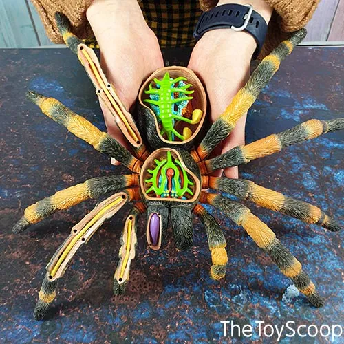
Once the prey is partially digested externally, the tarantula sucks up the liquefied nutrients through its mouth. The food then passes into the stomach, where further enzymatic digestion takes place. The digestive system then continues through the intestines, where nutrients are absorbed. The remaining indigestible waste is then eliminated. The stomach and intestines are essential for absorbing nutrients, allowing the spider to obtain the energy it needs to survive and thrive. The efficient digestive system helps the tarantula to maximize nutrient extraction from its prey, supporting its overall health.
Excretory System Waste Removal in Tarantulas
The excretory system in tarantulas removes metabolic waste products from the body, particularly nitrogenous waste. This is primarily managed by Malpighian tubules. These tubules filter hemolymph and extract waste, which is then processed and excreted, ensuring that the internal environment remains stable. The excretory system is critical for maintaining the overall health and function of the tarantula. This helps to prevent the buildup of toxic substances, ensuring the optimal functioning of other internal systems.
Malpighian Tubules Filtration and Excretion
Malpighian tubules are the primary excretory organs in tarantulas, functioning like kidneys. These tubules are located in the hemocoel and absorb waste products, such as uric acid, from the hemolymph. The waste is then transported to the hindgut, where it combines with feces and is excreted. This process is crucial for removing metabolic waste, maintaining osmotic balance, and ensuring the proper functioning of the internal systems. Malpighian tubules are critical for maintaining the tarantula’s internal balance and removing harmful waste products.
Nervous System Tarantulas’ Sensory Abilities
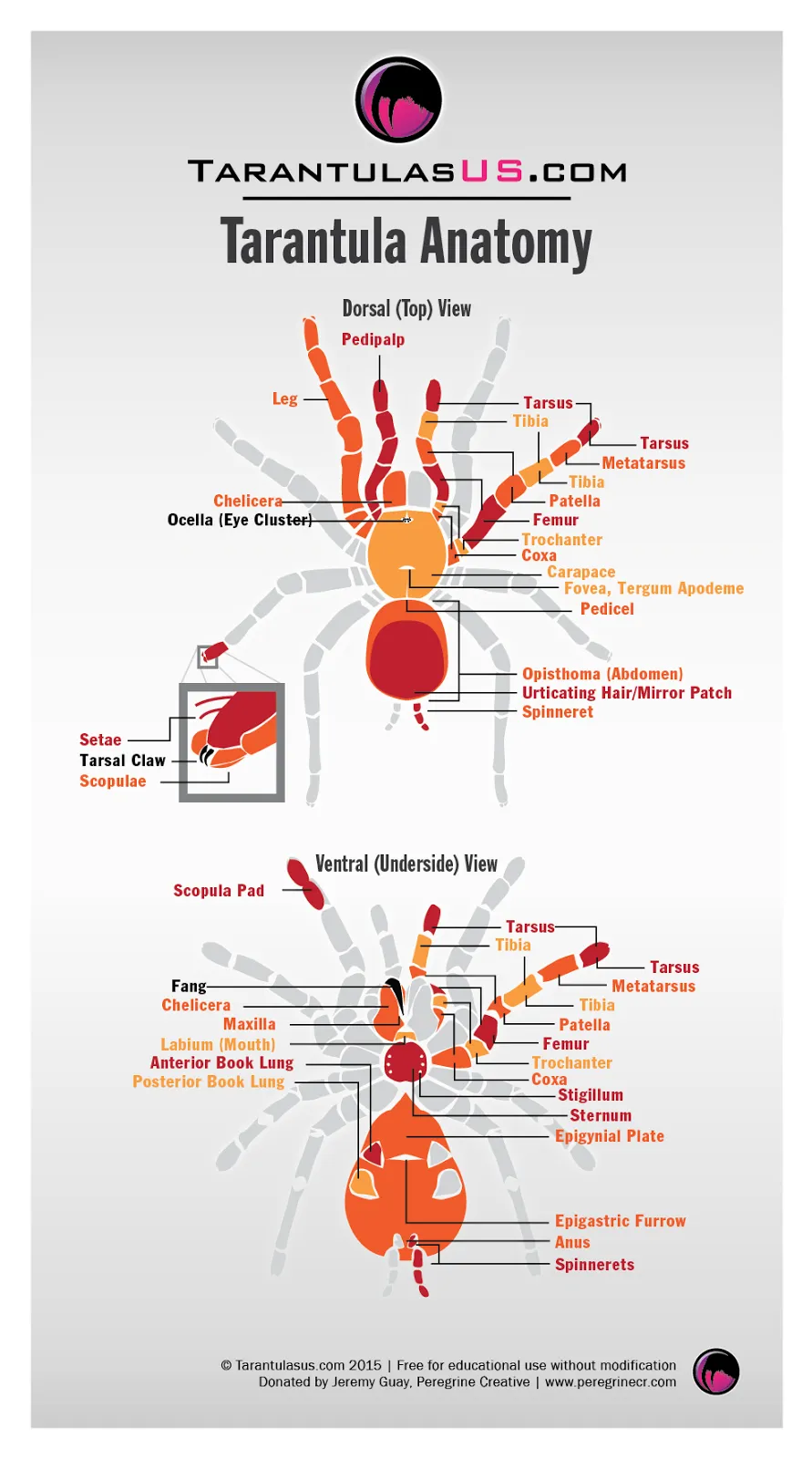
The nervous system of a tarantula allows for sensory perception, coordination, and behavior control. The brain, a cluster of ganglia, is located in the cephalothorax and processes sensory information. Nerves extend from the brain throughout the body, connecting to various sensory organs, such as the eyes, sensory hairs, and chemoreceptors. This comprehensive system is essential for hunting, avoiding danger, and navigating the environment. The tarantula’s nervous system is complex, enabling it to react to stimuli.
Brain and Nerve Structure
The tarantula brain is a centralized structure composed of multiple ganglia, the nerve centers that process sensory information and coordinate responses. Nerves radiate from the brain throughout the body, connecting to the various organs and sensory receptors. This complex network enables the tarantula to interpret stimuli, coordinate movement, and respond to its environment. The structure of the brain and nerves reflects the tarantula’s sophisticated sensory and behavioral capabilities. The brain and nerve structure control the tarantula’s movements and behavior.
Sensory Hairs and Their Function
Tarantulas are covered in sensory hairs (setae) that detect vibrations, air currents, and chemical signals. These hairs are extremely sensitive and play a crucial role in detecting prey, predators, and changes in the environment. The sensory hairs are connected to the nervous system and relay information to the brain, enabling the tarantula to react quickly to its surroundings. Sensory hairs are essential for the tarantula’s survival and are the primary way that it perceives the world. Sensory hairs enable the tarantula to detect vibrations, air currents, and chemical signals.
In conclusion, the organs of a tarantula are marvels of biological engineering, each with a specific role that contributes to the spider’s ability to survive and thrive. From the protective exoskeleton and efficient respiratory systems to the intricate digestive and nervous systems, every component is vital for the spider’s survival. Understanding how these organs function provides a deeper appreciation for the complexity and resilience of these fascinating creatures. Each organ plays a crucial role in ensuring the tarantula’s survival, making them well-adapted to their environment. This intricate interplay of organs allows tarantulas to adapt to their environment effectively, whether it is hunting prey or evading predators.
